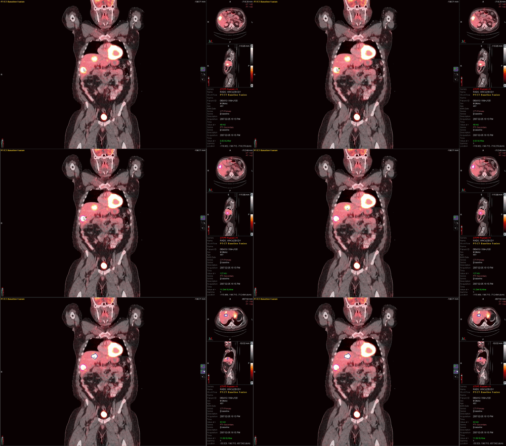
Quantitation is Needed to Predict Outcomes
SUVmax is a Useful Statistic, but it has Known Limitations
Many factors can influence SUVmax, such as its high dependence on noise, scan duration, and other acquisition parameters. These factors lead to poor reproducibility, and for this reason, you should be reluctant to rely solely on SUVmax for predicting outcomes. Quantitative metrics like metabolic tumor volume and total lesion glycolysis have demonstrated advantages over SUVmax alone1 and should be considered for inclusion in your reading workflow.
Referring Physicians Require Actionable Insights
Radiologists can't report effectively with ambiguous information. Extract crucial information beyond SUVmax alone to inform more confident treatment decisions.
Conventional Quantitative Metrics have Known Limitations
Due to the metric's known limitations2, you will experience situations where SUVmax cannot provide the information referring physicians require.
Time is of the Essence
Since no reimbursement options currently exist for capturing additional quantitative information, you will need a way to incorporate this information without spending any extra time.
MIM Encore®
An End-to-End Solution for Nuclear Medicine
MIM Encore provides a vendor-neutral solution for reconstruction, image processing, reading, and reporting.

Vendor Neutral
View and analyze PET and Nuclear Medicine images from virtually any camera manufacturer.
Increased Speed and Consistency
Custom reading workflows and image layouts are tailored to your preference. Link any number of studies automatically for easier comparison.
Advanced Therapy Response
LesionID® Pro and PET Edge®+ allow you to provide referring physicians with the latest clinically-significant staging and therapy response information, such as SUVmean, SUV Peak, and Total Metabolic Tumor Volume.
Increase Efficiency for the Nuclear Medicine Imaging Workflow by Implementing a Single Application.
SPECTRA Recon® supports SPECT/CT image reconstruction and CT attenuation correction for virtually any camera manufacturer. SPECTRA Quant® allows you to convert image counts to SUV.
MIM Encore provides a comprehensive suite of standard Nuclear Medicine processing tools, including lung quantification for SPECT/CT and liver functional analysis. Automated processing is available with MIM Assistant®.
Images can be accessed as a thin-client using floating licenses, or through a PACS plug-in. Patient data can be stored securely on the internet and accessed from anywhere from a workstation using MIM Zero Footprint™.
See How MIM Can Help You Imagine a New Reading Workflow
Schedule a Meeting Now